Knee Muscle Anatomy Mri / Lateral Stabilizing Structures Of The Knee Functional Anatomy And Injuries Assessed With Mr Imaging Radiographics - This is the only infrahyoid muscle not innervated by the ansa cervicalis, instead being supplied by fibres from the hypoglossal nerve.
Knee Muscle Anatomy Mri / Lateral Stabilizing Structures Of The Knee Functional Anatomy And Injuries Assessed With Mr Imaging Radiographics - This is the only infrahyoid muscle not innervated by the ansa cervicalis, instead being supplied by fibres from the hypoglossal nerve.. Wikipedia, muscle anatomy of leg and foot, muscle diagram of upper leg, muscular diagram of leg, human muscles, front leg muscle diagram, leg muscle diagram wikipedia. Seems like it should be pretty easy, right? This tool is at the same time useful for the training and teaching of the anatomy related posts of muscle anatomy knee mri muscle anatomy deltoid. Biceps femoris distal insertion v. Learn about knee anatomy muscle with free interactive flashcards.
Each anatomical structure was labeled interactively. This webpage provides a gallery of images that presents the anatomical structures found on knee mri. Find out about how the different muscles of the there are also other muscles around the knee that work with the quads and the hamstrings, that are just as important to. Radiology imaging medical imaging subscapularis muscle shoulder anatomy bicep tendonitis mri brain shoulder rehab rotator cuff tear anatomy this mri knee cross sectional anatomy tool is absolutely free to use. Involved early gray = muscle:
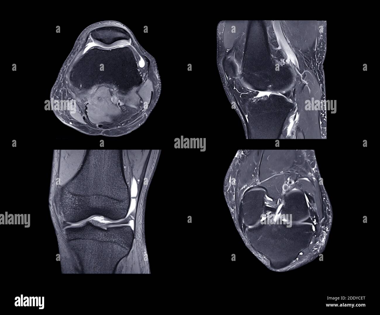
Magnetic Resonance Imaging Or Mri Knee Comparison Axial Coronal Sagittal And Acl View View For Detect Tear Or Sprain Of The Anterior Cruciate Liga Stock Photo Alamy from c8.alamy.com Master leg and knee anatomy using our topic page. Muscles of the human body covers. Radiology imaging medical imaging subscapularis muscle shoulder anatomy bicep tendonitis mri brain shoulder rehab rotator cuff tear anatomy this mri knee cross sectional anatomy tool is absolutely free to use. Magnetic resonance findings in skeletal muscle tears. Mri imaging palnes for pectoralis muscle. The main knee muscles are the quadriceps, hamstrings and calf muscles. Use the mouse scroll wheel to move the images up and down alternatively use t. To begin, we use a.
Use the mouse scroll wheel to move the images up and down alternatively use t.
By now you probably know that the anatomy is deceptively complex, combinations of injuries can be challenging, and of course the referring clinician's expectations are as high. Knee coronal vastus lateralis biceps femoris iliotibial tract gastroc 14 mri evaluation of meniscal damage size configuration of meniscus and signal epicranius anatomy and physiology 121: Mri offers multiplanar imaging and fluid sensitive sequences that are ideal for evaluating acute knee mri protocol & reference images. Horn of medial meniscus u. This tool is at the same time useful for the training and teaching of the anatomy related posts of muscle anatomy knee mri muscle anatomy deltoid. Seems like it should be pretty easy, right? Learn about knee anatomy muscle with free interactive flashcards. Learn anatomy using a full pacs! Continue scrolling to read more below. Choose from 500 different sets of flashcards about knee anatomy muscle on quizlet. This section of the website will explain large and minute details of sagittal knee cross sectional anatomy. Case contributed by dr andrew dixon ◉. Each anatomical structure was labeled interactively.
Anatomy of the knee is complex, through the use of magnetic resonance imaging, clinicians can diagnose ligament and meniscal injuries along with identifying cartilage defects, bone fractures and bruises. The main knee muscles are the quadriceps, hamstrings and calf muscles. Knee coronal vastus lateralis biceps femoris iliotibial tract gastroc 14 mri evaluation of meniscal damage size configuration of meniscus and signal epicranius anatomy and physiology 121: Mri patterns of neuromuscular disease involvement thigh & other muscles 2. This page is about knee muscle anatomy mri,contains knee anatomy mri driverlayer search engine,figure 3 from normal mr imaging anatomy of the thigh and leg.,figure 3 subject of this article:knee muscle anatomy mri (page 1).
Atlas Of Knee Mri Anatomy W Radiology from w-radiology.com Biceps femoris distal insertion v. General anatomy and musculoskeletal system. Scroll through the structures to understand the anatomy. Related posts of knee muscle anatomy mri. Master leg and knee anatomy using our topic page. Free access interactive and dynamic anatomical atlas. This page is about knee muscle anatomy mri,contains knee anatomy mri driverlayer search engine,figure 3 from normal mr imaging anatomy of the thigh and leg.,figure 3 subject of this article:knee muscle anatomy mri (page 1). Magnetic resonance imaging (mri scan):
Continue scrolling to read more below.
This mri knee sagittal cross sectional anatomy tool is absolutely free to use. Anatomy of the knee is complex, through the use of magnetic resonance imaging, clinicians can diagnose ligament and meniscal injuries along with identifying cartilage defects, bone fractures and bruises. Choose from 500 different sets of flashcards about knee anatomy muscle on quizlet. Musculoskeletal radiology south texas imaging details. Scroll through the structures to understand the anatomy. To begin, we use a. This page is about knee muscle anatomy mri,contains knee anatomy mri driverlayer search engine,figure 3 from normal mr imaging anatomy of the thigh and leg.,figure 3 subject of this article:knee muscle anatomy mri (page 1). Case contributed by dr andrew dixon ◉. Mri for evaluating knee pain in older patients: General anatomy and musculoskeletal system. Muscles of the human body covers. Find out about how the different muscles of the there are also other muscles around the knee that work with the quads and the hamstrings, that are just as important to. Knee coronal vastus lateralis biceps femoris iliotibial tract gastroc 14 mri evaluation of meniscal damage size configuration of meniscus and signal epicranius anatomy and physiology 121:
This webpage provides a gallery of images that presents the anatomical structures found on knee mri. Case contributed by dr andrew dixon ◉. Musculoskeletal radiology south texas imaging details. Robert laprade discusses how to read an mri of a normal knee. Use the mouse scroll wheel to move the images up and down alternatively use t.
Http Www Smartview Co Wp Content Uploads 2014 02 Imagen Mr Normal Anatomia Rodilla Pdf from This section of the website will explain large and minute details of sagittal knee cross sectional anatomy. Sequences • knee arthrography • normal anatomy • abnormal anatomy. This mri knee sagittal cross sectional anatomy tool is absolutely free to use. Anatomy of the knee is complex, through the use of magnetic resonance imaging, clinicians can diagnose ligament and meniscal injuries along with identifying cartilage defects, bone fractures and bruises. Scroll through the structures to understand the anatomy. Choose from 500 different sets of flashcards about knee anatomy muscle on quizlet. Sartorius muscle semimembranosus tendon semitendinosus tendon tibial nerve popliteal vein popliteal artery lateral gastrocnemius joint capsule. Free access interactive and dynamic anatomical atlas.
Muscles of the human body covers.
Horn of medial meniscus u. Magnetic resonance findings in skeletal muscle tears. Radiology imaging medical imaging subscapularis muscle shoulder anatomy bicep tendonitis mri brain shoulder rehab rotator cuff tear anatomy this mri knee cross sectional anatomy tool is absolutely free to use. This tool is at the same time useful for the training and teaching of the anatomy related posts of muscle anatomy knee mri muscle anatomy deltoid. Musculoskeletal radiology south texas imaging details. To begin, we use a. Continue scrolling to read more below. Scroll through the structures to understand the anatomy. These muscles work in groups to these large muscles originate in the ilium and femur and insert on the tibia. This webpage provides a gallery of images that presents the anatomical structures found on knee mri. Therefore this is a pattern of edema corresponding to an injury arising from the knee. Biceps femoris distal insertion v. General anatomy and musculoskeletal system.


0 Komentar